An Introduction to 3D imaging
Over the last years, 3D photography has become indispensable for face and body visualization, geometry and surgery planning. The use of quantitative methods to assess body and facial proportions as well as soft-tissue structure are becoming vital to clinics. Photo documentation and follow-up examinations all require objective measuring to be carried out in order to ensure outcome relevancy. 3D visualization and exploration of volumetric changes improve the way clinics view possible treatment areas.
The introduction of 3D technology has revolutionized clinics. Three-dimensional images improve visualization of skin structures otherwise difficult to view in two-dimensions. 3D images display a full illustration of the face and body and allow images to be viewed from multiple angles and at different zoom levels. Incorporating 3D images will completely eradicate the need for physicians to interpret dimension and volume from a 2D image alone. Today, the ability of a system to acquire standardized before/after images that objectively display treatment results is what is making 3D photography the industry-leading standard.
3D surface acquisition systems
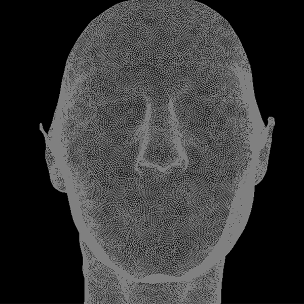
3D acquisition and reconstruction of the body can be done by laser scanner or stereophotogrammetry. Among the few techniques and devices that have been developed in the last decades, portable stereophotogrammetric systems have been proven to be the most efficient. For each image captured, two slightly offset photographs are taken in order to add depth and generate a single three-dimensional image, similar to the way the human eye sees. A 3D model is then obtained by a fusion of different shots; hence a face can be visualized from ear to ear and a body at 360°. The high resolution of images, coupled with the quantitative information embedded in the mesh, allow accuracy in body measurements. Furthermore, portable imaging devices have become more and more prevalent for clinics searching for image quality, speed and ease-of-use making portable devices the norm.
QuantifiCare technology choice
As pioneers in medical imaging, the QuantifiCare engineers have developed a stream-lined image capturing process combined with intuitive software package. The LifeViz®, portable and compact by design, are user-friendly systems that capture 3D images, making cumbersome 2D photography equipment redundant. We were the first to reconstruct 3D images of the body in 360°, revolutionary for a portable system. Standardization, accuracy and reliability are fundamental for effective image analysis and documentation. Our validated systems, used in clinical trials, are equipped with double flashes to ensure consistent lighting and good overall image reproducibility. They integrate dual-beam pointers for standardization in the capture process to enable accurate image comparison. Once images are acquired, a comprehensive software is needed. Our 3D software allows to quantify results, perform simulations and visualize pre- and post-operation results in 3D – capturing the synergy of multiple systems into one. The LifeViz® systems are poised to become an intrinsic part of any aesthetic practice.